VACANCIES                       CALL US: +91 98867 89799
FEEDBACK                     VACANCIES 
Best cornea surgery bagnalore
Cornea
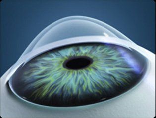
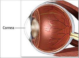
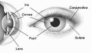
Like in a camera, the eye too has powerful and specially designed optics - the Cornea & Crystalline lens. These focus the image onto the retina. The Cornea does most of the focusing. It has no blood circulation and breaths oxygen from the air. This wonderful living ’lens’ is optically clear and is the most sensitive part of our body.

 Cornea Grafting Surgery At Karthik Netralaya Bangalore

The human eye and Cornea
The human eye is one of the most specialized organs of the body. Like in a camera, the eye too has powerful and specially designed optics - the Cornea & Crystalline lens. These focus the image onto the retina. The Cornea does most of the focusing. It has no blood circulation and breaths oxygen from the air. This wonderful living ’lens’ is optically clear and is the most sensitive part of our body.

Boman's membrane lies just beneath the epithelium. Because this layer is very tough and difficult to penetrate, it protects the cornea from injury.
The stroma is the thickest layer and lies just beneath Bowman's. It is composed of tiny collagen fibrils that run parallel to each other. This special formation of the collagen fibrils gives the cornea its clarity.

Descemet's membrane lies between the stroma and the endothelium. The endothelium is just underneath Descemet's and is only one cell layer thick. This layer pumps water from the cornea, keeping it clear. If damaged or disease, these cells will not regenerate.
Tiny vessels at the outermost edge of the cornea provide nourishment, along with the aqueous and tear film.

Corneal diseases
Corneal diseases can cause clouding and distortion of vision, and eventually blindness. There are many types of corneal diseases like infections due to contact lenses, dry eye, abrasions from trauma, and inflammations. Other conditions include keratoconus, pterygium etc.
Best cornea surgery bangalore
The layers of the cornea
The epithelium is layer of cells that cover the surface of the cornea. It is only about 5-6 cell layers thick and quickly regenerates when the cornea is injured. If the injury penetrates more deeply into the cornea, it may leave a scar. Scars leave opaque areas, causing the corneal to lose its clarity and luster.
Cornea
Like in a camera, the eye too has powerful and specially designed optics - the Cornea & Crystalline lens. These focus the image onto the retina. The Cornea does most of the focusing. It has no blood circulation and breaths oxygen from the air. This wonderful living ’lens’ is optically clear and is the most sensitive part of our body.

What is Cataract?

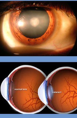
  • Cataract is the haziness of the crystalline lens of the eye. It causes scattering of light & blurring. It is not a layer that grows on the eye as many think.
  • Vision in dim or bright light may get affected. Headlights may cause glare at night. Spectacle power may change frequently. Day to day activities, working with gadgets, using stairs etc., will be increasingly difficult. Reading may improve initially.
  • It is best not to wait for cataract to mature as it can end up in complications. Except in special circumstances, a cataract surgery is performed when you cannot perform your day to day activities adequately and safely due to visual difficulties produced by the cataract. Many professions which demand very good eyesight may need an early surgery. However the natural lens, even with its slight imperfections of an early cataract, would have several advantages over the man made IOLs.
  • Cataract does not cause pain, headache, redness or watering. The vision loss in cataract is always slow, and if you ever have rapid or sudden decrease in vision, it should be informed, as you may need specialized investigations to exclude other eye diseases.
  • Cataract is common in middle age and in the elderly. It is also caused by drugs, chemicals, injuries, sun, UV light, radiation, electric shocks, malnutrition, diabetes, chronic diarrhea, X- rays, drugs etc. Viral infections during pregnancy, marriage within relatives, Genetic abnormalities etc. can cause cataracts in children.
Modern Micro-Surgery
 
Medicines, exercises, diet etc., cannot cure the cataract. Surgery is the only answer. It is the most often performed surgery on human beings.
  • Recent advancements have made cataract microsurgery safe & painless. The stay in the hospital is only few hours. Recovery is quick and one can go back to routine work in few days. The advanced cataract surgery here includes:
  • No injection, no pain, no bandage microsurgery
  • Self-healing, no stitch tunnel incisions
  • Central curvilinear capsulorhexis (CCC)
  • Excellent endothelial protection with Visco-procedures
  • Standard or premium (Aspherical, MICS, Toric & Multifocal) Intraocular lenses (IOLs)
  • Presbyopia correction
  • Safer surgeries for extremely complex & complicated situations
  • The nucleus of the cataract is cleared either by MICS, Phacoemulsification or Phacosection, using ultrasound or hydro-manual procedures. The actual technique is chosen based on the type of your cataract, with equitable safety & results. With this approach, all types from immature to hyper mature cataracts can be safely scientifically approached. Those with diabetes, hypertension, cardiac diseases, cancer & on anticoagulants (blood thinners), Cortico-steroids, anti-metabolites etc. can undergo these micro surgeries. Do not stop the blood thinners.
Karthik Netralaya offers you a very safe and advanced cataract microsurgery. Doctors are the first in Karnataka to introduce lOLs in 1981, ECCE in 1983, Phacoemulsification in 1991 & Phacosection in 1993. MICS was introduced in 1993 & Smart SMICS in 2016.

The Intraocular Lens (IOL)
 
IOLs have seen tremendous advancements ever since Sir Harold Ridley introduced them in 1959. Your spectacle power can also be corrected by titrating IOL power with Optical or Immersion biometry. Today there are several options and an IOL is chosen based upon your visual needs and budget. Standard IOLs: vision in both eyes are corrected for either distance (outdoor, sports, professional driving), near (reading and computer work), or intermediate (cooking, arts & craft, desk work, multi-tasking, indoor activities).
  • In Monovision correction the dominant eye is focused for distance and the other eye is focused for near or intermediate vision. This practical solution gives some spectacle independence.
  • Premium blue blocking IOLs for daylight outdoor activity.
  • Premium wavefront optimized asphercial IOLs for better contrast vision especially in dim light.
  • Premium Toric IOLs to correct astigmatism. Other options are LRI, ASA and LASIK.
  • Premium diffractive multifocal IOLs attempt to correct both distance and near sights, offering higher spectacle independence. They are today's solutions for Presbyopia. These IOLs cause some halos around lights and slightly lowered contrast sensitivity in dim light.
  • Smart MSICS is a new concept aimed at extending depth of field, and spectacle independency may be achieved to a certain range.
  • All IOLs block harmful UV light and protect the retina.
  • Karthik Netralaya offers a wide range of IOLs made in India & abroad, meeting your required criteria.
  • For those with very complex eye conditions, Bioptics with a combination of several techniques can be opted, to maximise possible visual recovery. Combination of Phacosurgery, LRI, Excimer LASER etc. could help in special situations.
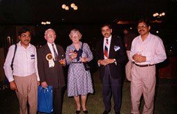
Dr. M S Ravindra with Prof. Harold Ridleyey
Excellent cost effective IOLs are now made in India. While the earlier IOLs were made of Poly-Methyl-Methacrylate (PMMA) and Silicone, today's foldable IOLs are made of Hydrophilic or Hydrophobic Acrylic material facilitating insertion through small incisions. Posterior Capsular Opacification (PCO) is less with Hydrophobic Acrylic IOLs.
  • ABOUT CORNEA

    The human eye and Cornea


    The human eye is one of the most specialized organs of the body. Like in a camera, the eye too has powerful and specially designed optics - the Cornea & Crystalline lens. These focus the image onto the retina. The Cornea does most of the focusing. It has no blood circulation and breaths oxygen from the air. This wonderful living ’lens’ is optically clear and is the most sensitive part of our body.


    The layers of the cornea


    The epithelium is layer of cells that cover the surface of the cornea. It is only about 5-6 cell layers thick and quickly regenerates when the cornea is injured. If the injury penetrates more deeply into the cornea, it may leave a scar. Scars leave opaque areas, causing the corneal to lose its clarity and luster.

    Boman's membrane lies just beneath the epithelium. Because this layer is very tough and difficult to penetrate, it protects the cornea from injury.

    The stroma is the thickest layer and lies just beneath Bowman's. It is composed of tiny collagen fibrils that run parallel to each other. This special formation of the collagen fibrils gives the cornea its clarity.

    Descemet's membrane lies between the stroma and the endothelium. The endothelium is just underneath Descemet's and is only one cell layer thick.  This layer pumps water from the cornea, keeping it clear. If damaged or disease, these cells will not regenerate.

    Tiny vessels at the outermost edge of the cornea provide nourishment, along with the aqueous and tear film.

  • KERATOCONUS

    What is Keratoconus?

    Keratoconus is a frequently seen corneal disease, occurring in about 1 in 1000 people, which typically starts after the age of 10 yrs. The hundreds of filaments of collagen layers in normal cornea are linked to each other by cross linking, giving it an enormous strength.  If these collagen cross-links are lost, as it happens in Keratoconus, there will be a progressive corneal thinning and stretching. This often occurs in both the eyes. The cornea bulges forward into an irregular cone shape.  This causes distortion of the image, which cannot be corrected with glasses. The eye develops irregular astigmatism (cylindrical errors) and myopia [shortsightedness] and the vision would become blurred.


    Who gets Keratoconus?

    Risk factors include eye rubbing, family history of keratoconus, genetic predisposition, certain systemic disorders such as Down’s syndrome, ocular allergy, connective tissue disease etc. Diabetics won’t develop Keratoconus because of natural crosslinking from high glucose and UV light. Affects men and women in equal proportions and is bilateral in 90% of cases.


    What are the symptoms?

    At early stages, the person feels the need for frequent change of spectacle correction. As the disease progresses, the vision deteriorates. Visual acuity becomes impaired at all distances, and night vision is sometimes quite poor. Keratoconus can cause substantial distortion of vision, with multiple images, streaking, sensitivity to light, eye strain from squinting in order to read, & itching in the eye.


    How it is diagnosed?

    This is usually done by an Ophthalmologist with a detailed eye examination including retinoscopy, keratometry, slit-lamp examination etc. Diagnosing early keratoconus can be tricky, since mild disease often does not show any signs on slit-lamp examination. Streak retinospcopy can pick up early Keratoconus.


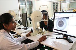
    At Karthik Netralaya a very sensitive instrument called the ‘Pentacam HR’ is used for early diagnosis. This is an automated instrument working on Shiemflug principal, and can pick up extremely early Keratocouns, that often starts in the back of the cornea.


    How Keratoconus is treated?

    Treatment of mild to moderate keratoconus is done temporarily by Contact lenses, which can be the regular RGP or specific and specialised, like Rose K lenses and mini scleral contact lenses depending upon the severity of the illness. The new modality of treatment is Corneal Collagen Crosslinking with Riboflavin (C3-R) stabilises the cornea from further deteriorating. In severe stages the person has to undergo corneal grafting surgery.

What is Cataract?

  • Cataract is the haziness of the crystalline lens of the eye. It causes scattering of light & blurring. It is not a layer that grows on the eye as many think.
  • Vision in dim or bright light may get affected. Headlights may cause glare at night. Spectacle power may change frequently. Day to day activities, working with gadgets, using stairs etc., will be increasingly difficult. Reading may improve initially.
  • It is best not to wait for cataract to mature as it can end up in complications. Except in special circumstances, a cataract surgery is performed when you cannot perform your day to day activities adequately and safely due to visual difficulties produced by the cataract. Many professions which demand very good eyesight may need an early surgery. However the natural lens, even with its slight imperfections of an early cataract, would have several advantages over the man made IOLs.
  • Cataract does not cause pain, headache, redness or watering. The vision loss in cataract is always slow, and if you ever have rapid or sudden decrease in vision, it should be informed, as you may need specialized investigations to exclude other eye diseases.
  • Cataract is common in middle age and in the elderly. It is also caused by drugs, chemicals, injuries, sun, UV light, radiation, electric shocks, malnutrition, diabetes, chronic diarrhea, X- rays, drugs etc. Viral infections during pregnancy, marriage within relatives, Genetic abnormalities etc. can cause cataracts in children.

Modern Micro-Surgery
 
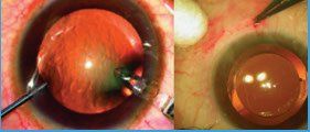
Medicines, exercises, diet etc., cannot cure the cataract. Surgery is the only answer. It is the most often performed surgery on human beings.
  • Recent advancements have made cataract microsurgery safe & painless. The stay in the hospital is only few hours. Recovery is quick and one can go back to routine work in few days. The advanced cataract surgery here includes:
  • No injection, no pain, no bandage microsurgery
  • Self-healing, no stitch tunnel incisions
  • Central curvilinear capsulorhexis (CCC)
  • Excellent endothelial protection with Visco-procedures
  • Standard or premium (Aspherical, MICS, Toric & Multifocal) Intraocular lenses (IOLs)
  • Presbyopia correction
  • Safer surgeries for extremely complex & complicated situations
  • The nucleus of the cataract is cleared either by MICS, Phacoemulsification or Phacosection, using ultrasound or hydro-manual procedures. The actual technique is chosen based on the type of your cataract, with equitable safety & results. With this approach, all types from immature to hyper mature cataracts can be safely scientifically approached. Those with diabetes, hypertension, cardiac diseases, cancer & on anticoagulants (blood thinners), Cortico-steroids, anti-metabolites etc. can undergo these micro surgeries. Do not stop the blood thinners.
Karthik Netralaya offers you a very safe and advanced cataract microsurgery. Doctors are the first in Karnataka to introduce lOLs in 1981, ECCE in 1983, Phacoemulsification in 1991 & Phacosection in 1993. MICS was introduced in 1993 & Smart SMICS in 2016.

The Intraocular Lens (IOL)
 
IOLs have seen tremendous advancements ever since Sir Harold Ridley introduced them in 1959. Your spectacle power can also be corrected by titrating IOL power with Optical or Immersion biometry. Today there are several options and an IOL is chosen based upon your visual needs and budget. Standard IOLs: vision in both eyes are corrected for either distance (outdoor, sports, professional driving), near (reading and computer work), or intermediate (cooking, arts & craft, desk work, multi-tasking, indoor activities).
  • In Monovision correction the dominant eye is focused for distance and the other eye is focused for near or intermediate vision. This practical solution gives some spectacle independence.
  • Premium blue blocking IOLs for daylight outdoor activity.
  • Premium wavefront optimized asphercial IOLs for better contrast vision especially in dim light.
  • Premium Toric IOLs to correct astigmatism. Other options are LRI, ASA and LASIK.
  • Premium diffractive multifocal IOLs attempt to correct both distance and near sights, offering higher spectacle independence. They are today's solutions for Presbyopia. These IOLs cause some halos around lights and slightly lowered contrast sensitivity in dim light.
  • Smart MSICS is a new concept aimed at extending depth of field, and spectacle independency may be achieved to a certain range.
  • All IOLs block harmful UV light and protect the retina.
  • Karthik Netralaya offers a wide range of IOLs made in India & abroad, meeting your required criteria.
  • For those with very complex eye conditions, Bioptics with a combination of several techniques can be opted, to maximise possible visual recovery. Combination of Phacosurgery, LRI, Excimer LASER etc. could help in special situations.
Dr. M S Ravindra with Prof. Harold Ridleyey
Excellent cost effective IOLs are now made in India. While the earlier IOLs were made of Poly-Methyl-Methacrylate (PMMA) and Silicone, today's foldable IOLs are made of Hydrophilic or Hydrophobic Acrylic material facilitating insertion through small incisions. Posterior Capsular Opacification (PCO) is less with Hydrophobic Acrylic IOLs.
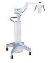
The new CXL technique & advanced Intacs XL Corneal cross linking equipment at Karthik Netralaya

What is collagen cross-linking?
A new modality of treatment for mild to moderate keratoconus in which riboflavin eye drops are applied to the eye. The Riboflavin, when activated for approximately 15-30 minutes with UV-A light, strengthens the collagen cross-links within the stroma and so recovers some of the cornea's mechanical strength. CSL or C3R has been shown to slow or arrest the progression of keratoconus, and in some cases even reverse it to an extent.

How does Cross Linking arrest Keratoconus?
This method works by increasing collagen cross-linking, which are the natural "anchors" within the cornea. These anchors are responsible for preventing the cornea from bulging out and becoming steep and irregular, which is the cause for keratoconus. Corneal collagen cross-linking (CXL) is established as an effective surgical treatment to increase the biomechanical stability of the cornea. We use CXL to treat idiopathic Keratoectasia such as keratoconus and pellucid marginal degeneration and post-LASIK ectasia etc.

The introduction of collagen cross linking (C3R, CXL, CCR, CCL and KXL) therapy promises to arrest the progression of conreal illensses. . The indications for CXL seem to be expanding dramatically, beyond conventionals. CXL is now being used preventively along with LASIK (called LASIK plus) and ASA (advanced surface ablations) in those who have risks of developing keratectasia as shown on Pentacam HR analysis. Conventional CXL machines cannot be used for these advanced treatments, and our new INTACS XL will be of enormous use in these special indications.

INTACS XL - A safe and effective CSL method to arrest progression of Keratoconus
It is a UVA collagen cross linking instrument designed with special focus on usage, effective performance and safety. It is a versatile UV irradiating device for performing a corneal cross-linking procedure at a wavelength at 365mn, designed with illumination intensity in the range of 3.0Mw/Cm to 30.Mw/Cm at a working distance of 50mm. While conventional machines take 30 minutes to deliver the UV dosage, this new machine delivers it in 3 minutes. This means quicker treatment, and better cooperation by the patient during the treatment.

What is Cataract?

  • Cataract is the haziness of the crystalline lens of the eye. It causes scattering of light & blurring. It is not a layer that grows on the eye as many think.
  • Vision in dim or bright light may get affected. Headlights may cause glare at night. Spectacle power may change frequently. Day to day activities, working with gadgets, using stairs etc., will be increasingly difficult. Reading may improve initially.
  • It is best not to wait for cataract to mature as it can end up in complications. Except in special circumstances, a cataract surgery is performed when you cannot perform your day to day activities adequately and safely due to visual difficulties produced by the cataract. Many professions which demand very good eyesight may need an early surgery. However the natural lens, even with its slight imperfections of an early cataract, would have several advantages over the man made IOLs.
  • Cataract does not cause pain, headache, redness or watering. The vision loss in cataract is always slow, and if you ever have rapid or sudden decrease in vision, it should be informed, as you may need specialized investigations to exclude other eye diseases.
  • Cataract is common in middle age and in the elderly. It is also caused by drugs, chemicals, injuries, sun, UV light, radiation, electric shocks, malnutrition, diabetes, chronic diarrhea, X- rays, drugs etc. Viral infections during pregnancy, marriage within relatives, Genetic abnormalities etc. can cause cataracts in children.

Modern Micro-Surgery
 
Medicines, exercises, diet etc., cannot cure the cataract. Surgery is the only answer. It is the most often performed surgery on human beings.
  • Recent advancements have made cataract microsurgery safe & painless. The stay in the hospital is only few hours. Recovery is quick and one can go back to routine work in few days. The advanced cataract surgery here includes:
  • No injection, no pain, no bandage microsurgery
  • Self-healing, no stitch tunnel incisions
  • Central curvilinear capsulorhexis (CCC)
  • Excellent endothelial protection with Visco-procedures
  • Standard or premium (Aspherical, MICS, Toric & Multifocal) Intraocular lenses (IOLs)
  • Presbyopia correction
  • Safer surgeries for extremely complex & complicated situations
  • The nucleus of the cataract is cleared either by MICS, Phacoemulsification or Phacosection, using ultrasound or hydro-manual procedures. The actual technique is chosen based on the type of your cataract, with equitable safety & results. With this approach, all types from immature to hyper mature cataracts can be safely scientifically approached. Those with diabetes, hypertension, cardiac diseases, cancer & on anticoagulants (blood thinners), Cortico-steroids, anti-metabolites etc. can undergo these micro surgeries. Do not stop the blood thinners.
Karthik Netralaya offers you a very safe and advanced cataract microsurgery. Doctors are the first in Karnataka to introduce lOLs in 1981, ECCE in 1983, Phacoemulsification in 1991 & Phacosection in 1993. MICS was introduced in 1993 & Smart SMICS in 2016.

The Intraocular Lens (IOL)
 
IOLs have seen tremendous advancements ever since Sir Harold Ridley introduced them in 1959. Your spectacle power can also be corrected by titrating IOL power with Optical or Immersion biometry. Today there are several options and an IOL is chosen based upon your visual needs and budget. Standard IOLs: vision in both eyes are corrected for either distance (outdoor, sports, professional driving), near (reading and computer work), or intermediate (cooking, arts & craft, desk work, multi-tasking, indoor activities).
  • In Monovision correction the dominant eye is focused for distance and the other eye is focused for near or intermediate vision. This practical solution gives some spectacle independence.
  • Premium blue blocking IOLs for daylight outdoor activity.
  • Premium wavefront optimized asphercial IOLs for better contrast vision especially in dim light.
  • Premium Toric IOLs to correct astigmatism. Other options are LRI, ASA and LASIK.
  • Premium diffractive multifocal IOLs attempt to correct both distance and near sights, offering higher spectacle independence. They are today's solutions for Presbyopia. These IOLs cause some halos around lights and slightly lowered contrast sensitivity in dim light.
  • Smart MSICS is a new concept aimed at extending depth of field, and spectacle independency may be achieved to a certain range.
  • All IOLs block harmful UV light and protect the retina.
  • Karthik Netralaya offers a wide range of IOLs made in India & abroad, meeting your required criteria.
  • For those with very complex eye conditions, Bioptics with a combination of several techniques can be opted, to maximise possible visual recovery. Combination of Phacosurgery, LRI, Excimer LASER etc. could help in special situations.
Dr. M S Ravindra with Prof. Harold Ridleyey
Excellent cost effective IOLs are now made in India. While the earlier IOLs were made of Poly-Methyl-Methacrylate (PMMA) and Silicone, today's foldable IOLs are made of Hydrophilic or Hydrophobic Acrylic material facilitating insertion through small incisions. Posterior Capsular Opacification (PCO) is less with Hydrophobic Acrylic IOLs.
  • DRY EYE

    What is Dry eye?

    The eye depends on the flow of tears to provide constant moisture and lubrication to maintain vision and comfort. Tears are a combination of water, for moisture; oils, for lubrication; mucus, for even spreading; and antibodies and special proteins, for resistance to infection. These components are secreted by special glands located around the eye. When there is an imbalance in this tear system, a person may experience dry eyes.


    What are the symptoms of dry eye?

    • Pain

    • Light sensitivity

    • A gritty sensation

    • A feeling of a foreign body or sand in the eye

    • Itching

    • Redness

    • Blurring of vision


    What Causes Dry Eyes?

    In addition to an imbalance in the tear-flow system of the eye, dry eyes can be caused by situations that dry out the tear film. This can be due to dry air from air conditioning, heat, or other environmental conditions.


    Karthik Netralaya proudly features "Oculus Dry Eye Lab" for the management of Dry Eye


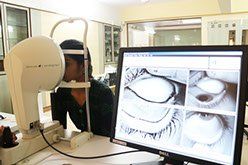
    This is a state-of-the-art technology for the comprehensive assessment of CVS, dry eye & tear film analysis and documentation. “Oculus Keratograph 5” offers several advanced diagnostic features in addition to its most reliable corneal surface topographic analysis. It incorporates a wide range of dry eye evaluation programs. Various parameters can be analysed quickly and non invasively.


    - Non invasive TBUT

    - Tear meniscus height

    - Meibomian gland analysis

    - Tear lipid analysis

    - Tear film dynamics study


    This new equipment revolutionises the diagnosis and management of dry eye & CVS especially in early stages. This is in addition to its diagnosing capabilities of corneal surface parameters in Keratoconus, pre LASIK & pre Cataract evaluations for premium IOL.

  • CORNEAL TRANSPLANTATION

    What is Corneal Transplantation?

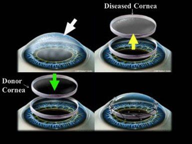
    Corneal Transplantation or Grafting is a fine micro surgery where a defective Cornea is replaced by a healthy donated Cornea. The Cornea of a departed donor is gifted by a humanitarian family and is collected & processed by an eye bank. Cornea grafting is a highly skilled day care surgery. In spite of enormous research, there is no synthetic equivalent to God given precious Cornea.


    When would one need a Corneal transplantation?

    Corneal transplantation is suggested when medicines fail to heal corneal diseases or when glasses or contact lenses cannot restore the vision due to corneal irregularities. Infections, Keratoconus, Injuries, scarring due to various reasons, corneal failure from previous surgeries, birth diseases etc. are some of the causes for corneal diseases.


    Cornea donors can be of any age, sex or blood group. Corneas are only collected from donors after death. Common health issues like Diabetes, Hypertension, Thyroid imbalances, use of spectacles etc. do not affect the health of the donor cornea. Corneas from persons having corneal diseases, advanced cancer, communicable diseases like HIV, Rabies, Hepatitis, Viral brain infections etc., are  not accepted.


    Types of Corneal transplantation


    • Penetrating Keratoplasty:

    Here the entire thickness of the diseased Cornea is replaced by a healthy donor Cornea & is secured by multiple tiny sutures. It takes a few months to years for the patient to get back his potential vision.


    • Deep Anterior Lamellar Keratoplasty: (DALK)

    Here the defective outer part of the Cornea is replaced, leaving behind the healthy inner Descemet’s and endothelial layer. The risk of graft rejection is  lesser in this procedure.


    • Descemet Stripping Endothelial Keratoplasty: (DSEK)

    Contrary to the previous surgery, only the unhealthy innermost layers of the Cornea are replaced with a thin layer donor Cornea, which includes some stroma Descemet’s and the healthy endothelium. This allows faster recovery of vision and better corneal  integrity.


     Descemet Membrane Endothelial Keratoplasty (DMEK)

    Here only the inner most ultra-thin single cell layered endothelium is transplanted along with its Descemet membrane. Your corneal surgeon will perform examinations like Slit lamp Biomicroscopy, Anterior segment OCT, Specular microscopy etc., and suggest the surgery best suited for you.

What is Cataract?

  • Cataract is the haziness of the crystalline lens of the eye. It causes scattering of light & blurring. It is not a layer that grows on the eye as many think.
  • Vision in dim or bright light may get affected. Headlights may cause glare at night. Spectacle power may change frequently. Day to day activities, working with gadgets, using stairs etc., will be increasingly difficult. Reading may improve initially.
  • It is best not to wait for cataract to mature as it can end up in complications. Except in special circumstances, a cataract surgery is performed when you cannot perform your day to day activities adequately and safely due to visual difficulties produced by the cataract. Many professions which demand very good eyesight may need an early surgery. However the natural lens, even with its slight imperfections of an early cataract, would have several advantages over the man made IOLs.
  • Cataract does not cause pain, headache, redness or watering. The vision loss in cataract is always slow, and if you ever have rapid or sudden decrease in vision, it should be informed, as you may need specialized investigations to exclude other eye diseases.
  • Cataract is common in middle age and in the elderly. It is also caused by drugs, chemicals, injuries, sun, UV light, radiation, electric shocks, malnutrition, diabetes, chronic diarrhea, X- rays, drugs etc. Viral infections during pregnancy, marriage within relatives, Genetic abnormalities etc. can cause cataracts in children.

Modern Micro-Surgery
 
Medicines, exercises, diet etc., cannot cure the cataract. Surgery is the only answer. It is the most often performed surgery on human beings.
  • Recent advancements have made cataract microsurgery safe & painless. The stay in the hospital is only few hours. Recovery is quick and one can go back to routine work in few days. The advanced cataract surgery here includes:
  • No injection, no pain, no bandage microsurgery
  • Self-healing, no stitch tunnel incisions
  • Central curvilinear capsulorhexis (CCC)
  • Excellent endothelial protection with Visco-procedures
  • Standard or premium (Aspherical, MICS, Toric & Multifocal) Intraocular lenses (IOLs)
  • Presbyopia correction
  • Safer surgeries for extremely complex & complicated situations
  • The nucleus of the cataract is cleared either by MICS, Phacoemulsification or Phacosection, using ultrasound or hydro-manual procedures. The actual technique is chosen based on the type of your cataract, with equitable safety & results. With this approach, all types from immature to hyper mature cataracts can be safely scientifically approached. Those with diabetes, hypertension, cardiac diseases, cancer & on anticoagulants (blood thinners), Cortico-steroids, anti-metabolites etc. can undergo these micro surgeries. Do not stop the blood thinners.
Karthik Netralaya offers you a very safe and advanced cataract microsurgery. Doctors are the first in Karnataka to introduce lOLs in 1981, ECCE in 1983, Phacoemulsification in 1991 & Phacosection in 1993. MICS was introduced in 1993 & Smart SMICS in 2016.

The Intraocular Lens (IOL)
 
IOLs have seen tremendous advancements ever since Sir Harold Ridley introduced them in 1959. Your spectacle power can also be corrected by titrating IOL power with Optical or Immersion biometry. Today there are several options and an IOL is chosen based upon your visual needs and budget. Standard IOLs: vision in both eyes are corrected for either distance (outdoor, sports, professional driving), near (reading and computer work), or intermediate (cooking, arts & craft, desk work, multi-tasking, indoor activities).
  • In Monovision correction the dominant eye is focused for distance and the other eye is focused for near or intermediate vision. This practical solution gives some spectacle independence.
  • Premium blue blocking IOLs for daylight outdoor activity.
  • Premium wavefront optimized asphercial IOLs for better contrast vision especially in dim light.
  • Premium Toric IOLs to correct astigmatism. Other options are LRI, ASA and LASIK.
  • Premium diffractive multifocal IOLs attempt to correct both distance and near sights, offering higher spectacle independence. They are today's solutions for Presbyopia. These IOLs cause some halos around lights and slightly lowered contrast sensitivity in dim light.
  • Smart MSICS is a new concept aimed at extending depth of field, and spectacle independency may be achieved to a certain range.
  • All IOLs block harmful UV light and protect the retina.
  • Karthik Netralaya offers a wide range of IOLs made in India & abroad, meeting your required criteria.
  • For those with very complex eye conditions, Bioptics with a combination of several techniques can be opted, to maximise possible visual recovery. Combination of Phacosurgery, LRI, Excimer LASER etc. could help in special situations.
Dr. M S Ravindra with Prof. Harold Ridleyey
Excellent cost effective IOLs are now made in India. While the earlier IOLs were made of Poly-Methyl-Methacrylate (PMMA) and Silicone, today's foldable IOLs are made of Hydrophilic or Hydrophobic Acrylic material facilitating insertion through small incisions. Posterior Capsular Opacification (PCO) is less with Hydrophobic Acrylic IOLs.
Corneal Surgeries at Karthik Netralaya
 
The department offers most recent advances in the field of corneal surgery which include:

• Penetratingkeratoplasty:
(Optical, tectonic and therapeutic) with anterior segment reconstruction, Combined with SFIOL Vitreo-retinal procedures and Paediatric PK

• Lamellar corneal surgeries:
Including Deep Anterior Lamellar Keratoplasty (DALK), Deep Lamellar Endothelial Keratoplasty (DLEK) and Descemet's Stripping EndoKeratoplasty (DSEK)

• Refractive corneal surgery:
Offers the state of art facility for various laser refractive procedures like LASIK, Epi-LASIK, ASA, PRK and Wavefront guided customized ablation
• Ocular surface reconstruction surgeries
• Collagen Cross Linking for keratoconus
 
Our corneal specialised equipment & facilities:
 
• Advanced Intacs XL Corneal cross linking equipment
• Oculus dry eye lab & Oculus Keratograph 5
• Specular microscopy
• Oculus Pentacam HR
• Corneal topography Tomey IV Topographer
• Spectral domain Corneal OCT
• Computerized Contact Lens module for Keratoconus contact lens fitting Low visual aids to enhance vision in those with uncorrectable vision
• Deep lamellar & endothelial (DALK & DSAEK) cornea penetrating keratoplasty
• ‘Automated Corneal Shaper’ (ACS) for lamellar corneal grafting surgeries
• Specialized Contact lens fitting – for irregular cornea, keratoconus and for therapeutic purposes
Best cornea surgery bangalore

Keratoconus

What is Keratoconus?
Keratoconus is a frequently seen corneal disease, occurring in about 1 in 1000 people, which typically starts after the age of 10 yrs. The hundreds of filaments of collagen layers in normal cornea are linked to each other by cross linking, giving it an enormous strength. If these collagen cross-links are lost, as it happens in Keratoconus, there will be a progressive corneal thinning and stretching. This often occurs in both the eyes. The cornea bulges forward into an irregular cone shape. This causes distortion of the image, which cannot be corrected with glasses. The eye develops irregular astigmatism (cylindrical errors) and myopia [shortsightedness] and the vision would become blurred.
Who gets Keratoconus?
Risk factors include eye rubbing, family history of keratoconus, genetic predisposition, certain systemic disorders such as Down’s syndrome, ocular allergy, connective tissue disease etc. Diabetics won’t develop Keratoconus because of natural crosslinking from high glucose and UV light. Affects men and women in equal proportions and is bilateral in 90% of cases.
What are the symptoms?
At early stages, the person feels the need for frequent change of spectacle correction. As the disease progresses, the vision deteriorates. Visual acuity becomes impaired at all distances, and night vision is sometimes quite poor. Keratoconus can cause substantial distortion of vision, with multiple images, streaking, sensitivity to light, eye strain from squinting in order to read, & itching in the eye.
How it is diagnosed?
This is usually done by an Ophthalmologist with a detailed eye examination including retinoscopy, keratometry, slit-lamp examination etc. Diagnosing early keratoconus can be tricky, since mild disease often does not show any signs on slit-lamp examination. Streak retinospcopy can pick up early Keratoconus.
At Karthik Netralaya a very sensitive instrument called the ‘Pentacam HR’ is used for early diagnosis. This is an automated instrument working on Shiemflug principal, and can pick up extremely early Keratocouns, that often starts in the back of the cornea.
How Keratoconus is treated?
Treatment of mild to moderate keratoconus is done temporarily by Contact lenses, which can be the regular RGP or specific and specialised, like Rose K lenses and mini scleral contact lenses depending upon the severity of the illness. The new modality of treatment is Corneal Collagen Crosslinking with Riboflavin (C3-R) stabilises the cornea from further deteriorating. In severe stages the person has to undergo corneal grafting surgery.
Best cornea surgery bangalore

The new CXL technique & advanced Intacs XL Corneal cross linking equipment at Karthik Netralaya

What is collagen cross-linking?
A new modality of treatment for mild to moderate keratoconus in which riboflavin eye drops are applied to the eye. The Riboflavin, when activated for approximately 15-30 minutes with UV-A light, strengthens the collagen cross-links within the stroma and so recovers some of the cornea's mechanical strength. CSL or C3R has been shown to slow or arrest the progression of keratoconus, and in some cases even reverse it to an extent.
How does Cross Linking arrest Keratoconus?
This method works by increasing collagen cross-linking, which are the natural "anchors" within the cornea. These anchors are responsible for preventing the cornea from bulging out and becoming steep and irregular, which is the cause for keratoconus. Corneal collagen cross-linking (CXL) is established as an effective surgical treatment to increase the biomechanical stability of the cornea. We use CXL to treat idiopathic Keratoectasia such as keratoconus and pellucid marginal degeneration and post-LASIK ectasia etc.
The introduction of collagen cross linking (C3R, CXL, CCR, CCL and KXL) therapy promises to arrest the progression of conreal illensses. . The indications for CXL seem to be expanding dramatically, beyond conventionals. CXL is now being used preventively along with LASIK (called LASIK plus) and ASA (advanced surface ablations) in those who have risks of developing keratectasia as shown on Pentacam HR analysis. Conventional CXL machines cannot be used for these advanced treatments, and our new INTACS XL will be of enormous use in these special indications. 
INTACS XL - A safe and effective CSL method to arrest progression of Keratoconus
It is a UVA collagen cross linking instrument designed with special focus on usage, effective performance and safety. It is a versatile UV irradiating device for performing a corneal cross-linking procedure at a wavelength at 365mn, designed with illumination intensity in the range of 3.0Mw/Cm to 30.Mw/Cm at a working distance of 50mm. While conventional machines take 30 minutes to deliver the UV dosage, this new machine delivers it in 3 minutes. This means quicker treatment, and better cooperation by the patient during the treatment.

Karthik Netralaya proudly features "Oculus Dry Eye Lab" for the management of Dry Eye
 
What is Dry eye?
The eye depends on the flow of tears to provide constant moisture and lubrication to maintain vision and comfort. Tears are a combination of water, for moisture; oils, for lubrication; mucus, for even spreading; and antibodies and special proteins, for resistance to infection. These components are secreted by special glands located around the eye. When there is an imbalance in this tear system, a person may experience dry eyes.

cornea eye hospital bangalore
This is a state-of-the-art technology for the comprehensive assessment of CVS, dry eye & tear film analysis and documentation. “Oculus Keratograph 5” offers several advanced diagnostic features in addition to its most reliable corneal surface topographic analysis. It incorporates a wide range of dry eye evaluation programs. Various parameters can be analysed quickly and non invasively.
  • Non invasive TBUT
  • Tear meniscus height
  • Meibomian gland analysis
  • Tear lipid analysis
  • Tear film dynamics study
This new equipment revolutionises the diagnosis and management of dry eye & CVS especially in early stages. This is in addition to its diagnosing capabilities of corneal surface parameters in Keratoconus, pre LASIK & pre Cataract evaluations for premium IOL.

cornea eye hospital bangalore

Corneal Transplantation

What is Corneal Transplantation?
Corneal Transplantation or Grafting is a fine micro surgery where a defective Cornea is replaced by a healthy donated Cornea. The Cornea of a departed donor is gifted by a humanitarian family and is collected & processed by an eye bank. Cornea grafting is a highly skilled day care surgery. In spite of enormous research, there is no synthetic equivalent to God given precious Cornea.

When would one need a Corneal transplantation?
Who gets Keratoconus?
Corneal transplantation is suggested when medicines fail to heal corneal diseases or when glasses or contact lenses cannot restore the vision due to corneal irregularities. Infections, Keratoconus, Injuries, scarring due to various reasons, corneal failure from previous surgeries, birth diseases etc. are some of the causes for corneal diseases.
Cornea donors can be of any age, sex or blood group. Corneas are only collected from donors after death. Common health issues like Diabetes, Hypertension, Thyroid imbalances, use of spectacles etc. do not affect the health of the donor cornea. Corneas from persons having corneal diseases, advanced cancer, communicable diseases like HIV, Rabies, Hepatitis, Viral brain infections etc., are not accepted.
Types of Corneal transplantation
  • Penetrating Keratoplasty:
Here the entire thickness of the diseased Cornea is replaced by a healthy donor Cornea & is secured by multiple tiny sutures. It takes a few months to years for the patient to get back his potential vision.
  • Deep Anterior Lamellar Keratoplasty: (DALK)
Here the defective outer part of the Cornea is replaced, leaving behind the healthy inner Descemet’s and endothelial layer. The risk of graft rejection is lesser in this procedure.
  • Descemet Stripping Endothelial Keratoplasty: (DSEK)
Contrary to the previous surgery, only the unhealthy innermost layers of the Cornea are replaced with a thin layer donor Cornea, which includes some stroma Descemet’s and the healthy endothelium. This allows faster recovery of vision and better corneal integrity cornea surgery bangalore, cornea transplant surgery, DALK, Karthik Netralaya.
  • Descemet Membrane Endothelial Keratoplasty (DMEK)
Here only the inner most ultra-thin single cell layered endothelium is transplanted along with its Descemet membrane. Your corneal surgeon will perform examinations like Slit lamp Biomicroscopy, Anterior segment OCT, Specular microscopy etc., and suggest the surgery best suited for you.

Corneal Surgeries at Karthik Netralaya
The department offers most recent advances in the field of corneal surgery which include:
• Penetratingkeratoplasty:
(Optical, tectonic and therapeutic) with anterior segment reconstruction, Combined with SFIOL Vitreo-retinal procedures and Paediatric PK
• Lamellar corneal surgeries:
Including Deep Anterior Lamellar Keratoplasty (DALK), Deep Lamellar Endothelial Keratoplasty (DLEK) and Descemet's Stripping EndoKeratoplasty (DSEK)
• Refractive corneal surgery:
Offers the state of art facility for various laser refractive procedures like LASIK, Epi-LASIK, ASA, PRK and Wavefront guided customized ablation
• Ocular surface reconstruction surgeries
• Collagen Cross Linking for keratoconus
 
Our corneal specialised equipment & facilities:
• Advanced Intacs XL Corneal cross linking equipment
• Oculus dry eye lab & Oculus Keratograph 5
• Specular microscopy
• Oculus Pentacam HR
• Corneal topography Tomey IV Topographer
• Spectral domain Corneal OCT
• Computerized Contact Lens module for Keratoconus contact lens fitting Low visual aids to enhance vision in those with uncorrectable vision
• Deep lamellar & endothelial (DALK & DSAEK) cornea penetrating keratoplasty
• ‘Automated Corneal Shaper’ (ACS) for lamellar corneal grafting surgeries
• Specialized Contact lens fitting – for irregular cornea, keratoconus and for therapeutic purposes

Share by: