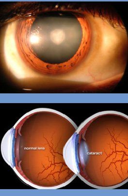
VACANCIES                       CALL US: +91 98867 89799
FEEDBACK                     VACANCIES 
Glaucoma eye care bangalore

Best glaucoma treatment karthik netralaya

Glaucoma
The Silent Thief of Sight !!
Glaucoma is a disease that causes damage to the nerve of the eye, the optic nerve, and gets worse over time if not recognised. It's often associated with an increase in eye pressure. Glaucoma tends to be inherited and may not show till mid age. The optic nerve transmits images to the brain. Permanent loss of vision can occur over several years, if glaucoma is not properly treated.

What is Cataract?

  • Cataract is the haziness of the crystalline lens of the eye. It causes scattering of light & blurring. It is not a layer that grows on the eye as many think.
  • Vision in dim or bright light may get affected. Headlights may cause glare at night. Spectacle power may change frequently. Day to day activities, working with gadgets, using stairs etc., will be increasingly difficult. Reading may improve initially.
  • It is best not to wait for cataract to mature as it can end up in complications. Except in special circumstances, a cataract surgery is performed when you cannot perform your day to day activities adequately and safely due to visual difficulties produced by the cataract. Many professions which demand very good eyesight may need an early surgery. However the natural lens, even with its slight imperfections of an early cataract, would have several advantages over the man made IOLs.
  • Cataract does not cause pain, headache, redness or watering. The vision loss in cataract is always slow, and if you ever have rapid or sudden decrease in vision, it should be informed, as you may need specialized investigations to exclude other eye diseases.
  • Cataract is common in middle age and in the elderly. It is also caused by drugs, chemicals, injuries, sun, UV light, radiation, electric shocks, malnutrition, diabetes, chronic diarrhea, X- rays, drugs etc. Viral infections during pregnancy, marriage within relatives, Genetic abnormalities etc. can cause cataracts in children.


Modern Micro-Surgery
 
Medicines, exercises, diet etc., cannot cure the cataract. Surgery is the only answer. It is the most often performed surgery on human beings.
  • Recent advancements have made cataract microsurgery safe & painless. The stay in the hospital is only few hours. Recovery is quick and one can go back to routine work in few days. The advanced cataract surgery here includes:
  • No injection, no pain, no bandage microsurgery
  • Self-healing, no stitch tunnel incisions
  • Central curvilinear capsulorhexis (CCC)
  • Excellent endothelial protection with Visco-procedures
  • Standard or premium (Aspherical, MICS, Toric & Multifocal) Intraocular lenses (IOLs)
  • Presbyopia correction
  • Safer surgeries for extremely complex & complicated situations
  • The nucleus of the cataract is cleared either by MICS, Phacoemulsification or Phacosection, using ultrasound or hydro-manual procedures. The actual technique is chosen based on the type of your cataract, with equitable safety & results. With this approach, all types from immature to hyper mature cataracts can be safely scientifically approached. Those with diabetes, hypertension, cardiac diseases, cancer & on anticoagulants (blood thinners), Cortico-steroids, anti-metabolites etc. can undergo these micro surgeries. Do not stop the blood thinners.
Karthik Netralaya offers you a very safe and advanced cataract microsurgery. Doctors are the first in Karnataka to introduce lOLs in 1981, ECCE in 1983, Phacoemulsification in 1991 & Phacosection in 1993. MICS was introduced in 1993 & Smart SMICS in 2016.

The Intraocular Lens (IOL)
 
IOLs have seen tremendous advancements ever since Sir Harold Ridley introduced them in 1959. Your spectacle power can also be corrected by titrating IOL power with Optical or Immersion biometry. Today there are several options and an IOL is chosen based upon your visual needs and budget. Standard IOLs: vision in both eyes are corrected for either distance (outdoor, sports, professional driving), near (reading and computer work), or intermediate (cooking, arts & craft, desk work, multi-tasking, indoor activities).
  • In Monovision correction the dominant eye is focused for distance and the other eye is focused for near or intermediate vision. This practical solution gives some spectacle independence.
  • Premium blue blocking IOLs for daylight outdoor activity.
  • Premium wavefront optimized asphercial IOLs for better contrast vision especially in dim light.
  • Premium Toric IOLs to correct astigmatism. Other options are LRI, ASA and LASIK.
  • Premium diffractive multifocal IOLs attempt to correct both distance and near sights, offering higher spectacle independence. They are today's solutions for Presbyopia. These IOLs cause some halos around lights and slightly lowered contrast sensitivity in dim light.
  • Smart MSICS is a new concept aimed at extending depth of field, and spectacle independency may be achieved to a certain range.
  • All IOLs block harmful UV light and protect the retina.
  • Karthik Netralaya offers a wide range of IOLs made in India & abroad, meeting your required criteria.
  • For those with very complex eye conditions, Bioptics with a combination of several techniques can be opted, to maximise possible visual recovery. Combination of Phacosurgery, LRI, Excimer LASER etc. could help in special situations.
Dr. M S Ravindra with Prof. Harold Ridleyey
Excellent cost effective IOLs are now made in India. While the earlier IOLs were made of Poly-Methyl-Methacrylate (PMMA) and Silicone, today's foldable IOLs are made of Hydrophilic or Hydrophobic Acrylic material facilitating insertion through small incisions. Posterior Capsular Opacification (PCO) is less with Hydrophobic Acrylic IOLs.
  • UNDERSTANDING GLAUCOMA

    Why Does Pressure Rise in the Eye to Cause Glaucoma?


    The pressure in the eye can increase when eye fluid isn't circulating normally in the front part of the eye.  This nourishing fluid, called aqueous humor, is produced by the Ciliary body, flows through pupil and than out of the eye through a mesh-like channel called Trabecular meshwork. If this channel gets blocked, fluid pressure builds up, causing glaucoma. There are several types of primary glaucoma. In the primary open angle glaucoma (POAG - the direct cause of the blockage is unknown, but doctors do know that it can passed from parents to children. In the primary closed angle glaucoma, decreased spaces and the crowding of structures in the front of the eye causes blockage at the pupil and the trabecular meshwork, causing sudden increase in the pressure.


    Other causes of glaucoma are a blunt or chemical injury to the eye, eye infections, blockage of retinal blood vessels, inflammatory conditions, and occasionally eye surgeries. Glaucoma usually occurs in both eyes, but may involve each eye to a different extent.


  • CLASSIFICATION OF GLAUCOMAS

    There are more than 40 types of glaucomas, but most fall into three principal categories; open angle, angle closure and congenital. These categories can be sub -classified as primary (occurring without a known cause) or secondary (due to an underlying cause, such as injury or illness).

    • Open angle glaucoma

    Open angle glaucoma is the most prevalent form of glaucoma and accounts for about 60% all glaucoma. In open angle glaucoma, the aqueous humor cannot get out through the trabecular meshwork.

    The onset of open angle glaucoma is slow with almost no symptoms. However, as time progresses, there is gradual loss of peripheral vision, a persistent elevation in IOP, optic-nerve atrophy, retinal ganglion cell atrophy etc. Blindness is the outcome unless surgery or drug treatment is initiated. Vision lost due to glaucoma is permanent and can never be recovered. Several conditions  are known to be associated with open angle glaucoma, like pigmentory dispersion syndrome, high myopia, retinal detachment, and diabetes mellitus etc.

    • Angle closure glaucoma

    Angle closure glaucoma is a  more severe form of glaucoma, and is seen more often in our country. The increase in IOP is related to sudden papillary blockage of aqueous humor circulation. This condition can be chronic (progressing slowly or occurring persistently) or acute (occurring suddenly). During an acute attack, the iris balloons forwardsand blocks the outflow channels suddenly and completely. IOP over 60 mmhg is not uncommon. It can lead to severe eye pain, headache, nausea, vomiting, stomach upsets, blurred vision accompanied by visions of colored halos around lights, and rapid loss of vision.

    • Secondary angle closure glaucoma

    Thisvmay develop as a result of a dislocated or swollen lens, post-surgery, Ciliary block or post-inflammatory conditions. Certain eye drops like atropine, pupil dilating medicines etc. can precipitate angle closure glaucoma in susceptible patients.

    • Congenital glaucomas

    Both subtypes congenital glaucoma—infant and juvenile- frequently require surgical intervention in preference to other, less effective medical treatments.

  • RISK & SYMPTOMS

    Who is at risk?

    Although anyone can get glaucoma, some people are at higher risk than others. They include:

    • Are over age 40
    • Have a family history of glaucoma
    • Have poor vision
    • Have diabetes
    • Take certain steroid medications, including steroid containing eye drops, ointments, tablets, skin ointments, inhalers, native & herbal medicines etc.
    • Have had trauma to the eye or eyes

    What are the symptoms of glaucoma? What happens when it is left untreated?


    At first, open angle glaucoma has no symptoms. Vision stays normal, and there is no pain. As glaucoma usually remains untreated for many years, people may notice that although they see things clearly in front of them, they miss objects to the side of their eye field of sight. At the end stages, it  may seem as though they are looking through a tunnel. Over time, the remaining forward vision may decrease until there is no vision left.


    If you have any of the following symptoms, seek immediate medical care:

    • Seeing halos around lights
    • Vision loss
    • Redness in the eye
    • Eye that looks hazy (particularly in infants)
    • Nausea or vomiting
    • Pain in the eye
    • Narrowing of vision (tunnel vision)

    How is glaucoma detected?

    Your Ophthalmologist can suspect glaucoma during  a routine eye test. Remember it's easy to manage glaucoma with your Ophthalmologist's advice and medications. It is also easy to miss Glaucoma, if you avoid consulting your Ophthalmologist. A spectacle check up or an eye screening in a camp, without a complete Ophthalmologists eye check up can easily miss the diagnosis.

  • DIAGNOSTIC TESTS

    Several procedures are used for diagnosing glaucoma. They, include the evaluation of IOP (tonometry), Optic nerve head and RNFL changes on slit lamp stereo biomicroscopy using high magnifying lens, visual field changes (perimetry), optic-disk analysis (OCT) and angle evaluation (gonioscopy).


    Tonometry

    Several  tonometry techniques can be used for the measurement of IOP. Applanation tonometry measures the force applied to the cornea per unit area. Air puff tonometry measures IOP by sending a "puff of air" onto the cornea to measure IOP. ICare tonometry measures the IOP by magnetic rebound method.


    Opthalmoscopy

    By this technique the ophthalmologist can directly evaluate the optic nerve using an biomicroscope or ophthalmoscope. Changes  in  morphology of the optic disk may point to glaucomatous damage.


    Perimetry

    This procedure tests for visual field defects, a defining feature of glaucomatous optic nerve damage. Isolated of impaired vision, surrounded by normal areas in a vision filed, are indicative of open angle glaucoma. Classical patterns are suggestive of glaucoma, but it is said that Perimetry becomes positive only after about 40 percent of ganglion cells are damaged!


    Gonioscopy

    This technique allows the ophthalmologist to view the anterior chamber angle directly. Both open angles and angle closure glaucomas need this test.

What is Cataract?

  • Cataract is the haziness of the crystalline lens of the eye. It causes scattering of light & blurring. It is not a layer that grows on the eye as many think.
  • Vision in dim or bright light may get affected. Headlights may cause glare at night. Spectacle power may change frequently. Day to day activities, working with gadgets, using stairs etc., will be increasingly difficult. Reading may improve initially.
  • It is best not to wait for cataract to mature as it can end up in complications. Except in special circumstances, a cataract surgery is performed when you cannot perform your day to day activities adequately and safely due to visual difficulties produced by the cataract. Many professions which demand very good eyesight may need an early surgery. However the natural lens, even with its slight imperfections of an early cataract, would have several advantages over the man made IOLs.
  • Cataract does not cause pain, headache, redness or watering. The vision loss in cataract is always slow, and if you ever have rapid or sudden decrease in vision, it should be informed, as you may need specialized investigations to exclude other eye diseases.
  • Cataract is common in middle age and in the elderly. It is also caused by drugs, chemicals, injuries, sun, UV light, radiation, electric shocks, malnutrition, diabetes, chronic diarrhea, X- rays, drugs etc. Viral infections during pregnancy, marriage within relatives, Genetic abnormalities etc. can cause cataracts in children.


Modern Micro-Surgery
 
Medicines, exercises, diet etc., cannot cure the cataract. Surgery is the only answer. It is the most often performed surgery on human beings.
  • Recent advancements have made cataract microsurgery safe & painless. The stay in the hospital is only few hours. Recovery is quick and one can go back to routine work in few days. The advanced cataract surgery here includes:
  • No injection, no pain, no bandage microsurgery
  • Self-healing, no stitch tunnel incisions
  • Central curvilinear capsulorhexis (CCC)
  • Excellent endothelial protection with Visco-procedures
  • Standard or premium (Aspherical, MICS, Toric & Multifocal) Intraocular lenses (IOLs)
  • Presbyopia correction
  • Safer surgeries for extremely complex & complicated situations
  • The nucleus of the cataract is cleared either by MICS, Phacoemulsification or Phacosection, using ultrasound or hydro-manual procedures. The actual technique is chosen based on the type of your cataract, with equitable safety & results. With this approach, all types from immature to hyper mature cataracts can be safely scientifically approached. Those with diabetes, hypertension, cardiac diseases, cancer & on anticoagulants (blood thinners), Cortico-steroids, anti-metabolites etc. can undergo these micro surgeries. Do not stop the blood thinners.
Karthik Netralaya offers you a very safe and advanced cataract microsurgery. Doctors are the first in Karnataka to introduce lOLs in 1981, ECCE in 1983, Phacoemulsification in 1991 & Phacosection in 1993. MICS was introduced in 1993 & Smart SMICS in 2016.

The Intraocular Lens (IOL)
 
IOLs have seen tremendous advancements ever since Sir Harold Ridley introduced them in 1959. Your spectacle power can also be corrected by titrating IOL power with Optical or Immersion biometry. Today there are several options and an IOL is chosen based upon your visual needs and budget. Standard IOLs: vision in both eyes are corrected for either distance (outdoor, sports, professional driving), near (reading and computer work), or intermediate (cooking, arts & craft, desk work, multi-tasking, indoor activities).
  • In Monovision correction the dominant eye is focused for distance and the other eye is focused for near or intermediate vision. This practical solution gives some spectacle independence.
  • Premium blue blocking IOLs for daylight outdoor activity.
  • Premium wavefront optimized asphercial IOLs for better contrast vision especially in dim light.
  • Premium Toric IOLs to correct astigmatism. Other options are LRI, ASA and LASIK.
  • Premium diffractive multifocal IOLs attempt to correct both distance and near sights, offering higher spectacle independence. They are today's solutions for Presbyopia. These IOLs cause some halos around lights and slightly lowered contrast sensitivity in dim light.
  • Smart MSICS is a new concept aimed at extending depth of field, and spectacle independency may be achieved to a certain range.
  • All IOLs block harmful UV light and protect the retina.
  • Karthik Netralaya offers a wide range of IOLs made in India & abroad, meeting your required criteria.
  • For those with very complex eye conditions, Bioptics with a combination of several techniques can be opted, to maximise possible visual recovery. Combination of Phacosurgery, LRI, Excimer LASER etc. could help in special situations.
Dr. M S Ravindra with Prof. Harold Ridleyey
Excellent cost effective IOLs are now made in India. While the earlier IOLs were made of Poly-Methyl-Methacrylate (PMMA) and Silicone, today's foldable IOLs are made of Hydrophilic or Hydrophobic Acrylic material facilitating insertion through small incisions. Posterior Capsular Opacification (PCO) is less with Hydrophobic Acrylic IOLs.
Optical Coherence Tomography (OCT) 

The leader in HD OCT, Optovue, at Karthik Netralaya is a modern spectral OCT equipment. It incorporates ocular coherence tomography technology. The Optovue OCT is the gold standard in vivo imaging device for glaucoma and offers proven reproducibility for diagnosis & follow ups. Very critical analysis of small changes in glaucoma damage is possible so that the treatment can be modulated. It has several unique objective glaucoma analysis modes. This includes RNFL thickness & TSNIT maps for nerve fiber layer analysis, GCC (Ganglion cell complex) mapping & analysis, and optic disc cup analysis.

Optovue OCT at Karthik Netralaya has updated normative database to compare patients test results. GCC analysis, one of the most recent criteria that can detect glaucoma in very early stages is extensively used. The OCT provides real-time cross-sectional images and quantitative analysis of glaucoma damage, and optimizes the diagnosis and monitoring of glaucoma defects.

What is Cataract?

  • Cataract is the haziness of the crystalline lens of the eye. It causes scattering of light & blurring. It is not a layer that grows on the eye as many think.
  • Vision in dim or bright light may get affected. Headlights may cause glare at night. Spectacle power may change frequently. Day to day activities, working with gadgets, using stairs etc., will be increasingly difficult. Reading may improve initially.
  • It is best not to wait for cataract to mature as it can end up in complications. Except in special circumstances, a cataract surgery is performed when you cannot perform your day to day activities adequately and safely due to visual difficulties produced by the cataract. Many professions which demand very good eyesight may need an early surgery. However the natural lens, even with its slight imperfections of an early cataract, would have several advantages over the man made IOLs.
  • Cataract does not cause pain, headache, redness or watering. The vision loss in cataract is always slow, and if you ever have rapid or sudden decrease in vision, it should be informed, as you may need specialized investigations to exclude other eye diseases.
  • Cataract is common in middle age and in the elderly. It is also caused by drugs, chemicals, injuries, sun, UV light, radiation, electric shocks, malnutrition, diabetes, chronic diarrhea, X- rays, drugs etc. Viral infections during pregnancy, marriage within relatives, Genetic abnormalities etc. can cause cataracts in children.


Modern Micro-Surgery
 
Medicines, exercises, diet etc., cannot cure the cataract. Surgery is the only answer. It is the most often performed surgery on human beings.
  • Recent advancements have made cataract microsurgery safe & painless. The stay in the hospital is only few hours. Recovery is quick and one can go back to routine work in few days. The advanced cataract surgery here includes:
  • No injection, no pain, no bandage microsurgery
  • Self-healing, no stitch tunnel incisions
  • Central curvilinear capsulorhexis (CCC)
  • Excellent endothelial protection with Visco-procedures
  • Standard or premium (Aspherical, MICS, Toric & Multifocal) Intraocular lenses (IOLs)
  • Presbyopia correction
  • Safer surgeries for extremely complex & complicated situations
  • The nucleus of the cataract is cleared either by MICS, Phacoemulsification or Phacosection, using ultrasound or hydro-manual procedures. The actual technique is chosen based on the type of your cataract, with equitable safety & results. With this approach, all types from immature to hyper mature cataracts can be safely scientifically approached. Those with diabetes, hypertension, cardiac diseases, cancer & on anticoagulants (blood thinners), Cortico-steroids, anti-metabolites etc. can undergo these micro surgeries. Do not stop the blood thinners.
Karthik Netralaya offers you a very safe and advanced cataract microsurgery. Doctors are the first in Karnataka to introduce lOLs in 1981, ECCE in 1983, Phacoemulsification in 1991 & Phacosection in 1993. MICS was introduced in 1993 & Smart SMICS in 2016.

The Intraocular Lens (IOL)
 
IOLs have seen tremendous advancements ever since Sir Harold Ridley introduced them in 1959. Your spectacle power can also be corrected by titrating IOL power with Optical or Immersion biometry. Today there are several options and an IOL is chosen based upon your visual needs and budget. Standard IOLs: vision in both eyes are corrected for either distance (outdoor, sports, professional driving), near (reading and computer work), or intermediate (cooking, arts & craft, desk work, multi-tasking, indoor activities).
  • In Monovision correction the dominant eye is focused for distance and the other eye is focused for near or intermediate vision. This practical solution gives some spectacle independence.
  • Premium blue blocking IOLs for daylight outdoor activity.
  • Premium wavefront optimized asphercial IOLs for better contrast vision especially in dim light.
  • Premium Toric IOLs to correct astigmatism. Other options are LRI, ASA and LASIK.
  • Premium diffractive multifocal IOLs attempt to correct both distance and near sights, offering higher spectacle independence. They are today's solutions for Presbyopia. These IOLs cause some halos around lights and slightly lowered contrast sensitivity in dim light.
  • Smart MSICS is a new concept aimed at extending depth of field, and spectacle independency may be achieved to a certain range.
  • All IOLs block harmful UV light and protect the retina.
  • Karthik Netralaya offers a wide range of IOLs made in India & abroad, meeting your required criteria.
  • For those with very complex eye conditions, Bioptics with a combination of several techniques can be opted, to maximise possible visual recovery. Combination of Phacosurgery, LRI, Excimer LASER etc. could help in special situations.
Dr. M S Ravindra with Prof. Harold Ridleyey
Excellent cost effective IOLs are now made in India. While the earlier IOLs were made of Poly-Methyl-Methacrylate (PMMA) and Silicone, today's foldable IOLs are made of Hydrophilic or Hydrophobic Acrylic material facilitating insertion through small incisions. Posterior Capsular Opacification (PCO) is less with Hydrophobic Acrylic IOLs.
  • TREATMENT

    How Is Glaucoma Treated?


    Glaucoma treatment may include prescription eye drops, LASER surgery, or microsurgery.


    Eye drops for glaucoma

    These either reduce the formation of fluid by the ciliary body or increase its outflow. Side effects of glaucoma drops may include allergy, redness of the eyes, itching, stinging, blurred vision or irritated eyes. Some glaucoma drugs may affect the heart and lungs. Be sure to tell your doctor about any other medications you are currently taking or are allergic to. If you have Asthma, cardiac diseases, hypertension or allergies, do mention to the doctor. Eye drops have to be used strictly as advised. Timings and dosages have to be maintained.

    Half hearted treatment leads to blindness, which cannot be reversed.  Attend to your follow ups as advised. Use the drops even on the date of your next visit. After instilling one drop, keep the eyes closed for 2 to 3 minutes, with the inner corners of the lids kept pressed, so that the eye drop is retained in the eyes, instead of draining into nose and throat. If there are any untoward effects after starting eye drops, do inform your doctor.


    LASER surgery for glaucoma

    LASER surgery for glaucoma (ALT or SLT) slightly increases the outflow of the fluid from the eye in open-angle glaucoma or eliminates fluid blockage in angle-closure glaucoma. Types of LASER surgery for glaucoma include Trabeculoplasty, in which a LASER is used to pull open the trabecular meshwork drainage area; iridotomy, in which a tiny hole is made in the iris, allowing the fluid to flow more freely; and cyclophotocoagulation, in which a laser beam treats ciliary body, reducing the production of fluid.


    Microsurgery for glaucoma

    In an operation called a Trabeculectomy, a new channel is created to drain the fluid, thereby reducing intraocular pressure. Sometimes this surgery fails due to the body's healing mechanisms closing the new channel, needing a redoing of the surgery. For some patients, a glaucoma implant is the best option. Other complications of microsurgery for glaucoma include temporary or permanent loss of vision, as well as bleeding or infection. 

  • TIPS FOR MANAGEMENT

    If you are diagnosed to have glaucoma, just do not worry. 

    • Continue your routine life style, with healthy modifications. Good diet, exercises, control of Diabetes, hypertension, weight & high cholesterol levels is important. Tell about your general health to the eye doctor and about your eye health to your physician.
    • Strictly follow your eye doctors advice. Use the medications regularly. Don’t alter them on your own, at any cost.
    • After putting one eye drop, press the inner corners of the closed eyes for 2 minutes.
    • Keep the drops in the fridge or a cool place so that you can use the drops beyond 1 month of its opening, Don’t touch the nozzle tip.
    • Use the drops as usual even on the day you visit Karthik Netralaya. Please don’t stop the drops, as the doctor needs to know the eye pressures while you are using the drops. Visit the doctor at different times so that the IOP gets checked around the clock. Usually the early morning IOP is the highest.
    • Visual fields & OCT are of paramount importance in the follow ups. You need to get them done atleast once an year.
    • Get your eye checked up before your birthday. It is the best way to remember your appointments. Avoid tight collars & neck tie. Avoid consuming large amount of fluids at any one time. Check if your skin ointment has any steroids.
    • Remember, the key to controlling glaucoma is catching it early. The best way to prevent vision loss form glaucoma is to know your risk factors and to have eye examinations at appropriate intervals.

What is Cataract?

  • Cataract is the haziness of the crystalline lens of the eye. It causes scattering of light & blurring. It is not a layer that grows on the eye as many think.
  • Vision in dim or bright light may get affected. Headlights may cause glare at night. Spectacle power may change frequently. Day to day activities, working with gadgets, using stairs etc., will be increasingly difficult. Reading may improve initially.
  • It is best not to wait for cataract to mature as it can end up in complications. Except in special circumstances, a cataract surgery is performed when you cannot perform your day to day activities adequately and safely due to visual difficulties produced by the cataract. Many professions which demand very good eyesight may need an early surgery. However the natural lens, even with its slight imperfections of an early cataract, would have several advantages over the man made IOLs.
  • Cataract does not cause pain, headache, redness or watering. The vision loss in cataract is always slow, and if you ever have rapid or sudden decrease in vision, it should be informed, as you may need specialized investigations to exclude other eye diseases.
  • Cataract is common in middle age and in the elderly. It is also caused by drugs, chemicals, injuries, sun, UV light, radiation, electric shocks, malnutrition, diabetes, chronic diarrhea, X- rays, drugs etc. Viral infections during pregnancy, marriage within relatives, Genetic abnormalities etc. can cause cataracts in children.


Modern Micro-Surgery
 
Medicines, exercises, diet etc., cannot cure the cataract. Surgery is the only answer. It is the most often performed surgery on human beings.
  • Recent advancements have made cataract microsurgery safe & painless. The stay in the hospital is only few hours. Recovery is quick and one can go back to routine work in few days. The advanced cataract surgery here includes:
  • No injection, no pain, no bandage microsurgery
  • Self-healing, no stitch tunnel incisions
  • Central curvilinear capsulorhexis (CCC)
  • Excellent endothelial protection with Visco-procedures
  • Standard or premium (Aspherical, MICS, Toric & Multifocal) Intraocular lenses (IOLs)
  • Presbyopia correction
  • Safer surgeries for extremely complex & complicated situations
  • The nucleus of the cataract is cleared either by MICS, Phacoemulsification or Phacosection, using ultrasound or hydro-manual procedures. The actual technique is chosen based on the type of your cataract, with equitable safety & results. With this approach, all types from immature to hyper mature cataracts can be safely scientifically approached. Those with diabetes, hypertension, cardiac diseases, cancer & on anticoagulants (blood thinners), Cortico-steroids, anti-metabolites etc. can undergo these micro surgeries. Do not stop the blood thinners.
Karthik Netralaya offers you a very safe and advanced cataract microsurgery. Doctors are the first in Karnataka to introduce lOLs in 1981, ECCE in 1983, Phacoemulsification in 1991 & Phacosection in 1993. MICS was introduced in 1993 & Smart SMICS in 2016.

The Intraocular Lens (IOL)
 
IOLs have seen tremendous advancements ever since Sir Harold Ridley introduced them in 1959. Your spectacle power can also be corrected by titrating IOL power with Optical or Immersion biometry. Today there are several options and an IOL is chosen based upon your visual needs and budget. Standard IOLs: vision in both eyes are corrected for either distance (outdoor, sports, professional driving), near (reading and computer work), or intermediate (cooking, arts & craft, desk work, multi-tasking, indoor activities).
  • In Monovision correction the dominant eye is focused for distance and the other eye is focused for near or intermediate vision. This practical solution gives some spectacle independence.
  • Premium blue blocking IOLs for daylight outdoor activity.
  • Premium wavefront optimized asphercial IOLs for better contrast vision especially in dim light.
  • Premium Toric IOLs to correct astigmatism. Other options are LRI, ASA and LASIK.
  • Premium diffractive multifocal IOLs attempt to correct both distance and near sights, offering higher spectacle independence. They are today's solutions for Presbyopia. These IOLs cause some halos around lights and slightly lowered contrast sensitivity in dim light.
  • Smart MSICS is a new concept aimed at extending depth of field, and spectacle independency may be achieved to a certain range.
  • All IOLs block harmful UV light and protect the retina.
  • Karthik Netralaya offers a wide range of IOLs made in India & abroad, meeting your required criteria.
  • For those with very complex eye conditions, Bioptics with a combination of several techniques can be opted, to maximise possible visual recovery. Combination of Phacosurgery, LRI, Excimer LASER etc. could help in special situations.
Dr. M S Ravindra with Prof. Harold Ridleyey
Excellent cost effective IOLs are now made in India. While the earlier IOLs were made of Poly-Methyl-Methacrylate (PMMA) and Silicone, today's foldable IOLs are made of Hydrophilic or Hydrophobic Acrylic material facilitating insertion through small incisions. Posterior Capsular Opacification (PCO) is less with Hydrophobic Acrylic IOLs.
Glaucoma is a disease that causes damage to the nerve of the eye, the optic nerve, and gets worse over time if not recognised. It's often associated with an increase in eye pressure. Glaucoma tends to be inherited and may not show till mid age. The optic nerve transmits images to the brain. Permanent loss of vision can occur over several years, if glaucoma is not properly treated.

Glaucoma Treatment at Karthik Netralaya Bangalore 
Why Does Pressure Rise in the Eye to Cause Glaucoma?
The pressure in the eye can increase when eye fluid isn't circulating normally in the front part of the eye. This nourishing fluid, called aqueous humor, is produced by the Ciliary body, flows through pupil and than out of the eye through a mesh-like channel called Trabecular meshwork. If this channel gets blocked, fluid pressure builds up, causing glaucoma. There are several types of primary glaucoma. In the primary open angle glaucoma (POAG - the direct cause of the blockage is unknown, but doctors do know that it can passed from parents to children. In the primary closed angle glaucoma, decreased spaces and the crowding of structures in the front of the eye causes blockage at the pupil and the trabecular meshwork, causing sudden increase in the pressure.

Other causes of glaucoma are a blunt or chemical injury to the eye, eye infections, blockage of retinal blood vessels, inflammatory conditions, and occasionally eye surgeries. Glaucoma usually occurs in both eyes, but may involve each eye to a different extent.
Glaucoma eye care bangalore
Classification of Glaucomas:

There are more than 40 types of glaucomas, but most fall into three principal categories; open angle, angle closure and congenital. These categories can be sub -classified as primary (occurring without a known cause) or secondary (due to an underlying cause, such as injury or illness).
  • Open angle glaucoma
Open angle glaucoma is the most prevalent form of glaucoma and accounts for about 60% all glaucoma. In open angle glaucoma, the aqueous humor cannot get out through the trabecular meshwork.
The onset of open angle glaucoma is slow with almost no symptoms. However, as time progresses, there is gradual loss of peripheral vision, a persistent elevation in IOP, optic-nerve atrophy, retinal ganglion cell atrophy etc. Blindness is the outcome unless surgery or drug treatment is initiated. Vision lost due to glaucoma is permanent and can never be recovered. Several conditions are known to be associated with open angle glaucoma, like pigmentory dispersion syndrome, high myopia, retinal detachment, and diabetes mellitus etc.
  • Angle closure glaucoma
Angle closure glaucoma is a more severe form of glaucoma, and is seen more often in our country. The increase in IOP is related to sudden papillary blockage of aqueous humor circulation. This condition can be chronic (progressing slowly or occurring persistently) or acute (occurring suddenly). During an acute attack, the iris balloons forwardsand blocks the outflow channels suddenly and completely. IOP over 60 mmhg is not uncommon. It can lead to severe eye pain, headache, nausea, vomiting, stomach upsets, blurred vision accompanied by visions of colored halos around lights, and rapid loss of vision.
  • Secondary angle closure glaucoma
Thisvmay develop as a result of a dislocated or swollen lens, post-surgery, Ciliary block or post-inflammatory conditions. Certain eye drops like atropine, pupil dilating medicines etc. can precipitate angle closure glaucoma in susceptible patients.
  • Congenital glaucomas
Both subtypes congenital glaucoma—infant and juvenile- frequently require surgical intervention in preference to other, less effective medical treatments.

Who is at risk?
Although anyone can get glaucoma, some people are at higher risk than others. They include :
  • Are over age 40
  • Have a family history of glaucoma
  • Have poor vision
  • Have diabetes
  • Take certain steroid medications, including steroid containing eye drops, ointments, tablets, skin ointments, inhalers, native & herbal medicines etc.
  • Have had trauma to the eye or eyes
Glaucoma eye surgery bangalore

What are the symptoms of glaucoma? What happens when it is left untreated?

At first, open angle glaucoma has no symptoms. Vision stays normal, and there is no pain. As glaucoma usually remains untreated for many years, people may notice that although they see things clearly in front of them, they miss objects to the side of their eye field of sight. At the end stages, it may seem as though they are looking through a tunnel. Over time, the remaining forward vision may decrease until there is no vision left.
If you have any of the following symptoms, seek immediate medical care:
  • Seeing halos around lights
  • Vision loss
  • Redness in the eye
  • Eye that looks hazy (particularly in infants)
  • Nausea or vomiting
  • Pain in the eye
  • Narrowing of vision (tunnel vision)
Best glaucoma care bangalore
How is glaucoma detected?
Your Ophthalmologist can suspect glaucoma during a routine eye test. Remember it's easy to manage glaucoma with your Ophthalmologist's advice and medications. It is also easy to miss Glaucoma, if you avoid consulting your Ophthalmologist. A spectacle check up or an eye screening in a camp, without a complete Ophthalmologists eye check up can easily miss the diagnosis.

Diagnostic tests for glaucoma
Several procedures are used for diagnosing glaucoma. They, include the evaluation of IOP (tonometry), Optic nerve head and RNFL changes on slit lamp stereo biomicroscopy using high magnifying lens, visual field changes (perimetry), optic-disk analysis (OCT) and angle evaluation (gonioscopy).
Tonometry
Several tonometry techniques can be used for the measurement of IOP. Applanation tonometry measures the force applied to the cornea per unit area. Air puff tonometry measures IOP by sending a "puff of air" onto the cornea to measure IOP. ICare tonometry measures the IOP by magnetic rebound method.
Opthalmoscopy
By this technique the ophthalmologist can directly evaluate the optic nerve using an biomicroscope or ophthalmoscope. Changes in morphology of the optic disk may point to glaucomatous damage.
Perimetry
This procedure tests for visual field defects, a defining feature of glaucomatous optic nerve damage. Isolated of impaired vision, surrounded by normal areas in a vision filed, are indicative of open angle glaucoma. Classical patterns are suggestive of glaucoma, but it is said that Perimetry becomes positive only after about 40 percent of ganglion cells are damaged!
Glaucoma eye hospital bangalore
Gonioscopy
This technique allows the ophthalmologist to view the anterior chamber angle directly. Both open angles and angle closure glaucomas need this test.

Optical Coherence Tomography (OCT)
The leader in HD OCT, Optovue, at Karthik Netralaya is a modern spectral OCT equipment. It incorporates ocular coherence tomography technology. The Optovue OCT is the gold standard in vivo imaging device for glaucoma and offers proven reproducibility for diagnosis & follow ups. Very critical analysis of small changes in glaucoma damage is possible so that the treatment can be modulated. It has several unique objective glaucoma analysis modes. This includes RNFL thickness & TSNIT maps for nerve fiber layer analysis, GCC (Ganglion cell complex) mapping & analysis, and optic disc cup analysis.
Optovue OCT at Karthik Netralaya has updated normative database to compare patients test results. GCC analysis, one of the most recent criteria that can detect glaucoma in very early stages is extensively used. The OCT provides real-time cross-sectional images and quantitative analysis of glaucoma damage, and optimizes the diagnosis and monitoring of glaucoma defects.
How Is Glaucoma Treated?
Glaucoma treatment may include prescription eye drops, LASER surgery, or microsurgery.
Eye drops for glaucoma
These either reduce the formation of fluid by the ciliary body or increase its outflow. Side effects of glaucoma drops may include allergy, redness of the eyes, itching, stinging, blurred vision or irritated eyes. Some glaucoma drugs may affect the heart and lungs. Be sure to tell your doctor about any other medications you are currently taking or are allergic to. If you have Asthma, cardiac diseases, hypertension or allergies, do mention to the doctor. Eye drops have to be used strictly as advised. Timings and dosages have to be maintained. Half hearted treatment leads to blindness, which cannot be reversed. Attend to your follow ups as advised. Use the drops even on the date of your next visit. After instilling one drop, keep the eyes closed for 2 to 3 minutes, with the inner corners of the lids kept pressed, so that the eye drop is retained in the eyes, instead of draining into nose and throat. If there are any untoward effects after starting eye drops, do inform your doctor.

LASER surgery for glaucoma
LASER surgery for glaucoma (ALT or SLT) slightly increases the outflow of the fluid from the eye in open-angle glaucoma or eliminates fluid blockage in angle-closure glaucoma. Types of LASER surgery for glaucoma include Trabeculoplasty, in which a LASER is used to pull open the trabecular meshwork drainage area; iridotomy, in which a tiny hole is made in the iris, allowing the fluid to flow more freely; and cyclophotocoagulation, in which a laser beam treats ciliary body, reducing the production of fluid.
best hospital for glaucoma bangalore
Microsurgery for glaucoma
In an operation called a Trabeculectomy, a new channel is created to drain the fluid, thereby reducing intraocular pressure. Sometimes this surgery fails due to the body's healing mechanisms closing the new channel, needing a redoing of the surgery. For some patients, a glaucoma implant is the best option. Other complications of microsurgery for glaucoma include temporary or permanent loss of vision, as well as bleeding or infection.

Tips for Glaucoma management

If you are diagnosed to have glaucoma, just do not worry.
  • Continue your routine life style, with healthy modifications. Good diet, exercises, control of Diabetes, hypertension, weight & high cholesterol levels is important. Tell about your general health to the eye doctor and about your eye health to your physician.
  • Strictly follow your eye doctors advice. Use the medications regularly. Don’t alter them on your own, at any cost.
  • After putting one eye drop, press the inner corners of the closed eyes for 2 minutes.
  • Keep the drops in the fridge or a cool place so that you can use the drops beyond 1 month of its opening, Don’t touch the nozzle tip. 
  • Use the drops as usual even on the day you visit Karthik Netralaya. Please don’t stop the drops, as the doctor needs to know the eye pressures while you are using the drops. Visit the doctor at different times so that the IOP gets checked around the clock. Usually the early morning IOP is the highest.
  • Visual fields & OCT are of paramount importance in the follow ups. You need to get them done atleast once an year.
  • Get your eye checked up before your birthday. It is the best way to remember your appointments. Avoid tight collars & neck tie. Avoid consuming large amount of fluids at any one time. Check if your skin ointment has any steroids.
  • Remember, the key to controlling glaucoma is catching it early. The best way to prevent vision loss form glaucoma is to know your risk factors and to have eye examinations at appropriate intervals.
Share by: